Tumor-suppressor genes
If oncogenes are a stuck accelerator, tumor-suppressor genes are the brakes and the repair crew. Their proteins keep cancer from starting, so the danger comes when their function is lost.
Two jobs that prevent cancer
The proteins made by tumor-suppressor genes usually do one of two jobs. Some maintain the genome’s integrity: they watch for DNA damage, repair it, or stop a damaged cell from dividing. Others are negative regulators of cell division, proteins that hold division back until the cell is supposed to divide.
| Gene | Job | Protein’s role |
|---|---|---|
| p53 | Genome integrity | Senses DNA damage, then triggers repair, stops the cell cycle, or causes apoptosis |
| BRCA-1, BRCA-2 | Genome integrity | Sense DNA damage and help repair it; mutated in some inherited breast cancers |
| DNA repair genes | Genome integrity | Fix damaged DNA; one is defective in people with xeroderma pigmentosum |
| Rb | Negative regulation | Blocks the genes a cell needs to copy its DNA and divide |
| NF1 | Negative regulation | Makes Ras hydrolyze its GTP, switching Ras off |
Losing a genome-integrity gene doesn’t make a cell divide by itself. It lets mutations pile up faster, including the mutations that create oncogenes. Losing a negative regulator speeds division directly.
Check yourselfLosing the NF1 protein has the same effect as which oncogene mutation?
A ras mutation that blocks GTP hydrolysis. Without NF1, Ras stays in its active, GTP-bound form, so the growth signal stays on and cell division speeds up.
Checkpoints in the cell cycle
A dividing cell goes through a repeating series of stages called the cell cycle. In G₁ the cell grows, in S it copies its DNA, in G₂ it prepares to divide, and in M it divides. A cell moves from one stage to the next when proteins called cyclins bind and switch on enzymes called cyclin-dependent kinases (cdks).
At several points the cycle can be halted. A checkpoint protein checks the genome for damage and, if it finds a problem, stops the cell from moving past that point, often by preventing cyclin–cdk complexes from becoming active. Checkpoint proteins aren’t needed for healthy cells to divide. Their job is to stop division when something is wrong, so a cell carrying a mutation doesn’t pass it to two daughter cells.
p53, the guardian at G₁
p53 is a checkpoint protein and a transcription factor. When DNA is damaged, the cell makes more p53, and p53 switches on genes whose proteins do two things: stop the cell cycle at the G₁ checkpoint, before the DNA is copied in S phase, and repair the DNA. If the repair succeeds, the cell continues through the cycle.
If the damage is too severe to repair, p53 activates genes that trigger apoptosis, the programmed cell death you met in Chapter 9. Killing an occasional cell that might turn cancerous is a good trade for a multicellular organism.
About half of all human cancers, including many tumors of the lung, breast, liver, and brain, carry mutations in p53.
Check yourselfCells lacking p53 divide normally in a dish, but after a dose of X-rays they keep dividing while normal cells pause. Why?
p53 isn’t needed for ordinary division, only for responding to DNA damage. Without it, damaged cells aren’t stopped at the G₁ checkpoint, repaired, or killed by apoptosis.
Rb holds back E2F
The Rb protein is a negative regulator of cell division. It is named for retinoblastoma, a cancer of the eye’s retina that develops when both copies of the Rb gene are lost.
Rb works by controlling E2F, a transcription factor that turns on the genes a cell needs to pass from G₁ into S phase. As long as Rb is bound to E2F, E2F can’t do its job and the cell can’t divide.
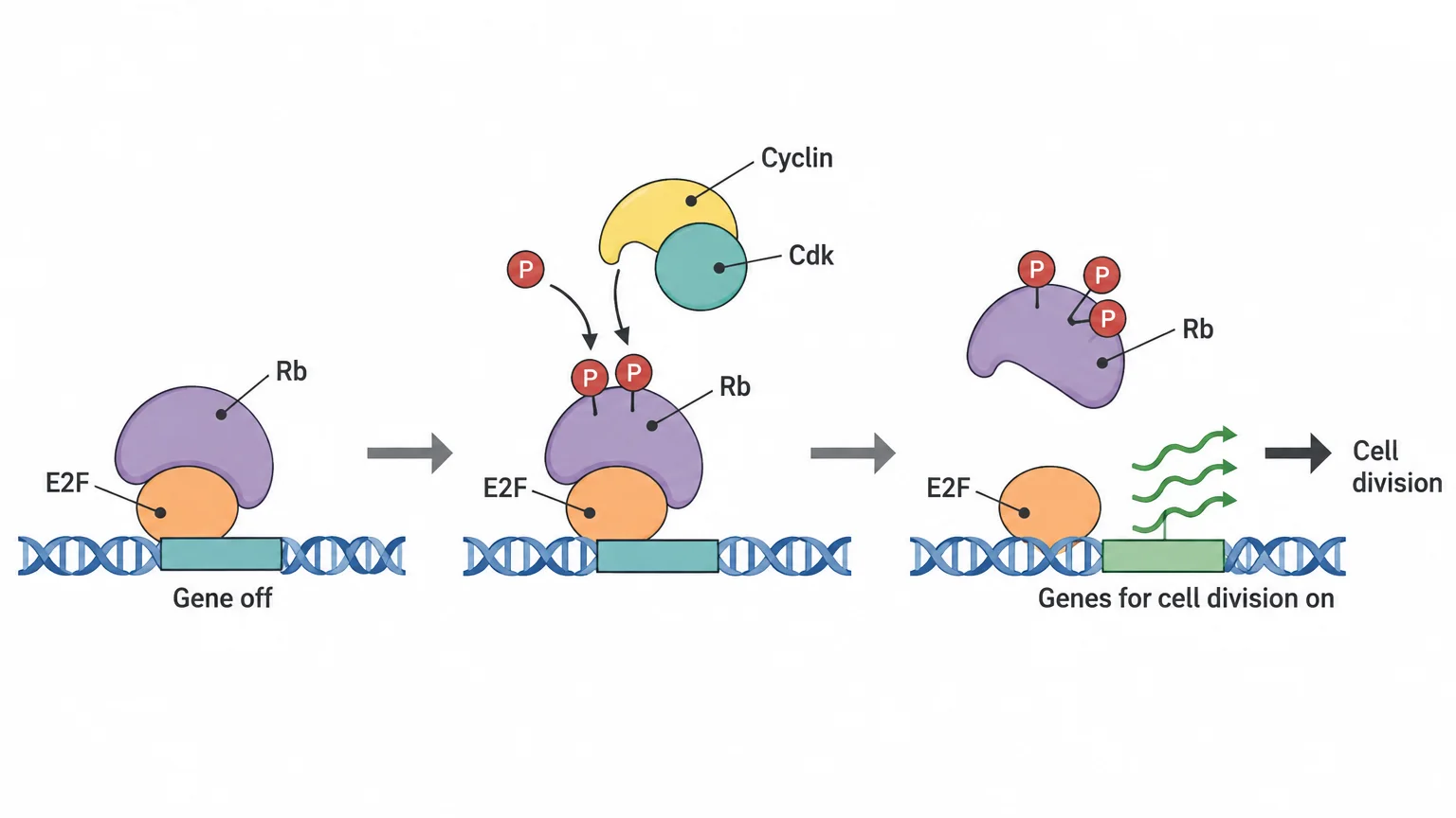
If both copies of Rb are defective, nothing holds E2F back. E2F stays active all the time, and the cells divide uncontrollably, as they do in retinoblastoma.
Practice questions
1 of 3
When a healthy cell is ready to divide, what releases E2F from the Rb protein?
2 of 3
Losing a tumor-suppressor gene that repairs DNA doesn’t make a cell divide faster by itself. How does it still promote cancer?
3 of 3
A protein inspects the genome for damage and, if it finds a problem, stops the cell from moving to the next stage of the cell cycle. What kind of protein is this?