Oncogenes
Healthy cells divide only when told to. In Chapter 9 you followed the signal: a growth factor binds its receptor, a chain of relay proteins and kinases passes the message inward, and transcription factors switch on genes that drive division. Many oncogenes are broken parts of exactly this kind of pathway.
Oncogenes keep the growth signal on
A growth factor pathway has three main kinds of proteins, and a mutation that makes any of them overactive can turn its gene into an oncogene.
| Protein | Normal job |
|---|---|
| Growth factor receptor | Binds the growth factor and starts the signal, such as the EGF receptor |
| Intracellular signaling protein | Relays the signal inside the cell, such as Ras |
| Transcription factor | Turns on the genes that make the cell divide |
Normally each of these proteins is active only while a growth factor is present. An oncogene keeps one of them switched on all the time, so the pathway tells the cell to divide even when no signal has arrived.
Ras is the textbook case. Recall that Ras is active while it holds GTP and turns itself off by hydrolyzing GTP to GDP. Mutations that make Ras an oncogene either slow that hydrolysis or make Ras swap GDP for GTP faster. Either way, more Ras sits in its active form, and the cell keeps getting the message to divide.
Proto-oncogenes
Oncogenes don’t come from nowhere. They start as ordinary, useful genes. A proto-oncogene is a normal gene that can become an oncogene if it is mutated. The normal ras gene is a proto-oncogene; a mutated, overactive ras is an oncogene.
Once mutated, the gene is expressed too much, expressed in the wrong kind of cell, or makes a protein that is too active. Four kinds of genetic change commonly do this.
| Change | What happens | Result |
|---|---|---|
| Missense mutation | One amino acid in the protein changes | The protein becomes overactive |
| Gene amplification | The gene is copied many extra times in the chromosome | Too much of the protein is made |
| Chromosomal translocation | A piece of one chromosome attaches to a different chromosome | A fused gene makes an overactive fusion protein |
| Retroviral insertion | A virus inserts its DNA next to the proto-oncogene | The viral promoter drives overexpression |
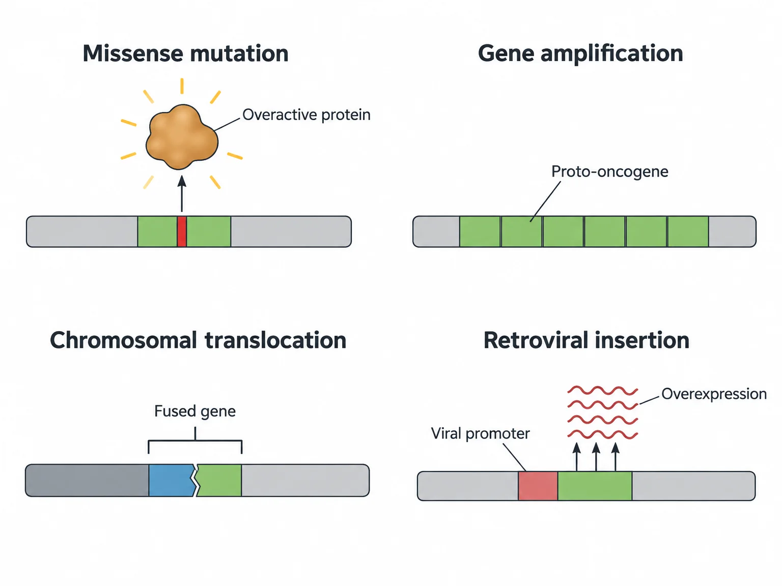
The ras gene is the classic missense example: a single amino acid change weakens Ras’s ability to hydrolyze its GTP, so Ras stays switched on.
A gene amplification is an abnormal increase in the number of copies of a gene. More copies mean more mRNA and more protein. Many human cancers, including some breast cancers, carry amplified proto-oncogenes.
Check yourselfA tumor’s cells have 30 copies of a proto-oncogene instead of the normal two. Which kind of change is this, and why does it promote cancer?
Gene amplification. The extra copies produce far too much of the protein, which overstimulates cell division even though the protein itself is normal.
Chromosomal translocations
In a chromosomal translocation, a segment of one chromosome breaks off and joins a different chromosome. If the break falls inside genes, the rejoined pieces can fuse two genes into one.
In one form of leukemia, a cancer of white blood cells, two chromosomes swap pieces. The promoter and first part of a gene called bcr are joined to part of a proto-oncogene called abl. The fused gene codes a fusion protein that is overactive.
The type of cancer follows from the promoter. The bcr promoter is normally active in white blood cells, so the fused gene is expressed there, and the result is a cancer of white blood cells.
Check yourselfIn this leukemia, why is the fusion protein made in white blood cells and not, say, in skin cells?
The fused gene is controlled by the bcr promoter, which is active in white blood cells. A promoter determines which cells transcribe the gene, so the overactive protein appears where bcr is normally expressed.
Viruses that cause cancer
Most cancers come from mutagens, but a few viruses cause cancer in animals and plants. In humans, the hepatitis B virus can cause liver cancer. Viruses do it in two ways.
The first is retroviral insertion. A retrovirus is a kind of virus that inserts a DNA copy of its genes into the host cell’s chromosome. Viral genes have strong promoters built for high expression. If the viral DNA happens to land beside a proto-oncogene, the viral promoter can drive that gene to be overexpressed.
The second is carrying an oncogene. One virus that causes sarcomas, tumors of connective tissue such as bone or cartilage, carries a gene called src. Normal cells have their own src gene, a proto-oncogene, which the virus picked up long ago. In the virus, src is transcribed from a powerful viral promoter, so infected cells make far too much Src protein and divide uncontrollably.
Practice questions
1 of 2
Some breast cancer cells carry many extra copies of a proto-oncogene whose coding sequence is perfectly normal. Why does this promote cancer?
2 of 2
A virus that causes sarcomas carries a gene that ordinary cells also have as a proto-oncogene. Why does the viral copy cause cancer?