Tight junctions and gap junctions
Anchoring junctions hold cells together, but they don’t stop things from slipping through the cracks between cells, and they don’t let cells share their contents. Animals use two other junctions for those jobs: one seals, the other connects.
Tight junctions seal a cell layer
The lining of your intestine is a sheet just one cell thick. One side of each cell faces the lumen (the hollow inside of the intestine, where food passes), and the other side faces the ECM and a blood vessel. If material could seep between the cells, the contents of your gut would leak straight into your blood, and blood components would leak out into your gut.
Tight junctions prevent that. They form a tight seal between neighboring cells so material can’t leak across the cell layer between them.
The seal is made of two membrane-spanning proteins, occludin and claudin. In each cell, they form strands woven through the plasma membrane like a net. The strands of one cell bind to the strands of the next, zipping the two membranes together.
Tight junctions are not mechanically strong, because they lack firm connections to the cytoskeleton. So cells joined by tight junctions also have anchoring junctions to hold them in place.
Three jobs of tight junctions
- Prevent leaks. In the intestine, they stop materials from passing between the lumen and the blood in either direction.
- Keep the cell’s two sides different. Membrane proteins can drift sideways, and tight junctions act as a fence that stops them. The side facing the lumen is the apical side; the side facing the blood is the basolateral side. Proteins for taking material in (receptor-mediated endocytosis) stay apical, and proteins for releasing it (exocytosis) stay basolateral. This difference between the sides is cell polarity. It lets intestinal cells absorb nutrients from the gut and release them into the blood, moving material across the cell layer, which is called transepithelial transport.
- Block microbes. Skin and the lining of the digestive tract are sealed by tight junctions. Some disease-causing microbes, including some that cause diarrhea, infect by breaking these seals.
Testing the seal
Researchers showed how well tight junctions work by injecting a rat’s bloodstream with lanthanum, a metal that shows up dark under the electron microscope. The lanthanum seeped into the spaces between intestinal cells on the blood side, but stopped dead at the tight junction and never reached the lumen side.
Gap junctions connect cells directly
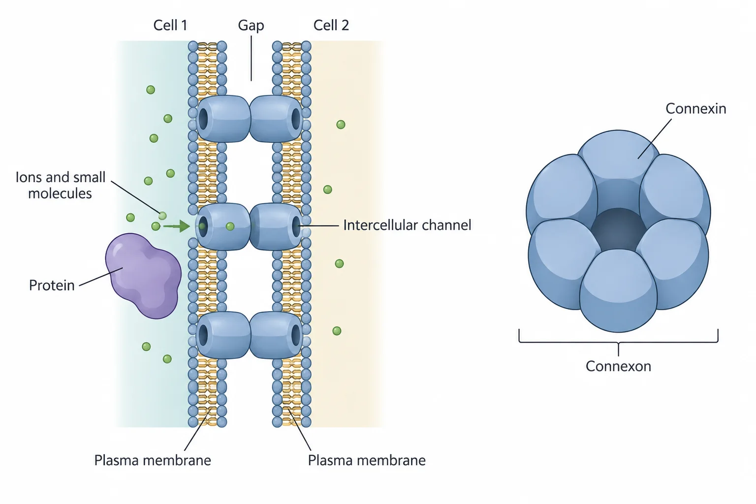
A gap junction is named for the small gap left between the plasma membranes of the two cells it connects. Gap junctions are clusters of tiny tunnels that join the cytosols of neighboring cells, so small things can pass straight from one cell to the next.
The tunnels are built from a membrane protein. In vertebrates it is connexin; invertebrates use a similar protein called innexin.
- Six connexin proteins in one cell’s plasma membrane form a ring with a channel in the middle. This ring is a connexon.
- A connexon in one cell lines up with a connexon in the neighboring cell, forming one continuous channel between the two cells.
- Many of these paired connexons cluster together in the membrane. That cluster is the gap junction.
What fits through
Gap junction channels pass ions such as Ca²⁺ and small molecules such as amino acids, sugars, and signaling molecules like cAMP. They are too small for RNA, proteins, or polysaccharides. So connected cells can share small metabolites and signal each other directly, yet each still keeps its own set of large molecules.
The size limit is usually stated in daltons (Da), a unit of mass for molecules; one dalton is about the mass of a hydrogen atom.
Researchers found the cutoff with glowing dyes. They grew a single layer of rat liver cells, which formed gap junctions with each other, and injected one cell with a fluorescent dye of known mass. Under a fluorescence microscope, they then checked whether the glow spread into the neighboring cells. Repeating this with dyes of different masses gave a clear pattern:
Later work in other cell types and species found some variation, but molecules heavier than about 1000 Da generally don’t pass.
Why the heart needs them
Gap junctions are abundant wherever cells must act together. Heart muscle cells are joined by many of them. Because ions flow through, an electrical change in one heart cell spreads quickly to its neighbors, so the cells contract together as a coordinated beat.
Check yourselfWhat would go wrong if the cells lining your small intestine had no tight junctions?
Practice questions
1 of 4
In intestinal lining cells, tight junctions keep nutrient-uptake proteins on the side facing the gut and export proteins on the side facing the blood. What does this arrangement make possible?
2 of 4
A diarrhea-causing microbe breaks down the tight junctions in a rat’s intestinal lining. Lanthanum, which shows up dark under the electron microscope, is then injected into the rat’s blood. Where in the lining would the lanthanum most likely appear?
3 of 4
A researcher injects one cell in a sheet of cultured liver cells that are joined by gap junctions. Which injected molecule would most likely spread into the neighboring cells?
4 of 4
In a dye-transfer experiment, single cells in a layer of cultured cells joined by gap junctions were injected with dyes of different masses. Dyes of 450, 600, and 850 Da spread into neighboring cells; dyes of 1100, 1500, and 2000 Da never did. Which conclusion do these data best support?