Cell junctions and the proteins that anchor cells
The ECM and cell wall give a body strength, but cells also need to connect directly to their neighbors. They do it with cell junctions, specialized structures where cells make physical contact with each other or with the ECM.
Five kinds of junction
Animal cells lack a rigid wall, so they rely on a wider variety of junctions than plant cells do.
| Organism | Junction | What it does |
|---|---|---|
| Animals | Anchoring junction | Holds cells to each other or to the ECM; mechanically strong |
| Animals | Tight junction | Seals neighboring cells in a layer so material can’t leak between them |
| Animals | Gap junction | A cluster of channels that let ions and small molecules pass directly from one cell’s cytosol to the next |
| Plants | Middle lamella | A polysaccharide layer that cements neighboring cell walls together |
| Plants | Plasmodesmata | Openable passageways through the cell walls that link the cytosols of neighboring cells |
Anchoring junctions
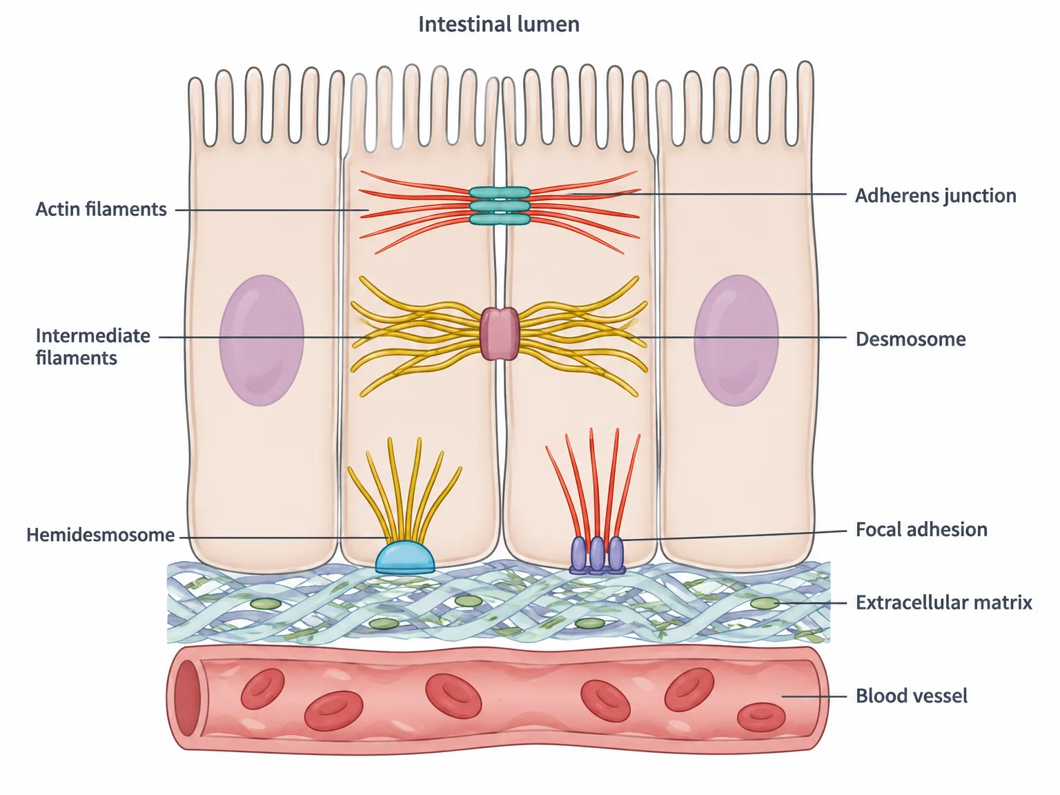
Anchoring junctions attach cells to each other and to the ECM. They are common wherever cells are tightly packed into linings. The single layer of cells lining the small intestine is a good example: anchoring junctions keep these cells stuck together, forming a strong barrier between the inside of the intestine and the blood.
The actual connections are made by cell adhesion molecules, or CAMs. These are proteins that span the plasma membrane: one end reaches outside to grip a partner, and the other end reaches into the cell. Inside the cell, CAMs attach to the cytoskeleton, the cell’s internal network of protein filaments. Two kinds of filament are involved:
- Actin filaments, thin filaments involved in cell shape and movement.
- Intermediate filaments, ropelike filaments that resist tension.
Linking the CAMs to the cytoskeleton is what makes anchoring junctions strong: the sturdy internal frameworks of neighboring cells end up tied together.
Four types of anchoring junction
The four types differ in two ways: what they attach to (another cell or the ECM) and which filament they connect to inside the cell.
| Junction | Attaches cell to | CAM used | Filament inside the cell |
|---|---|---|---|
| Adherens junction | Another cell (often as a band around the cell) | Cadherin | Actin filaments |
| Desmosome | Another cell (spotlike rivets) | Cadherin | Intermediate filaments |
| Hemidesmosome | The ECM | Integrin | Intermediate filaments |
| Focal adhesion | The ECM | Integrin | Actin filaments |
Cadherins: cell-to-cell glue
Cadherins are CAMs that link one cell to another. The outer part of a cadherin in one cell binds the outer part of a cadherin in the neighboring cell. This binding needs calcium ions (Ca²⁺), which change the cadherin’s shape so the two can lock together. The name comes from this: Ca²⁺-dependent adhering molecule. Inside the cell, linker proteins connect cadherins to actin or intermediate filaments.
Cadherins come in several varieties, such as E-cadherin and N-cadherin, and they bind like-to-like. E-cadherin binds only E-cadherin on the neighboring cell; it won’t bind N-cadherin. This like-to-like pairing is called homophilic binding (“homo” = same, “philic” = loving).
Integrins: cell-to-ECM glue
Integrins are CAMs that link a cell to the ECM. Unlike cadherins, they do not need Ca²⁺. Each integrin is built from two different subunits. Its outer part binds ECM components such as fibronectin (which in turn binds collagen fibers), and its inner part binds, through linker proteins, to actin or intermediate filaments. The result is a strong chain from the cytoskeleton, through the membrane, to the matrix.
More than mechanical
CAMs were first seen as simple fasteners, but they also take part in cell communication. Forming or breaking an anchoring junction changes signal transduction pathways (the chains of proteins that relay signals inside a cell), and those pathways can in turn change CAMs and loosen or tighten junctions.
CAMs also keep cells where they belong. When cancer-causing mutations make CAMs defective, cells lose their grip on their neighbors and the ECM. They can then break away and spread to other parts of the body, which is called metastasis. A mutation that inactivates a cadherin or an integrin would therefore be expected to promote metastasis.
Check yourselfHow are cadherins and integrins alike, and how do they differ?
Both are membrane-spanning cell adhesion molecules that connect to actin or intermediate filaments inside the cell, and both take part in cell signaling. Cadherins bind cells to other cells and require Ca²⁺; integrins bind cells to the ECM and do not need Ca²⁺.
Practice questions
1 of 2
Cadherins and integrins were first thought to act only as fasteners. What else have experiments shown they do?
2 of 2
Lung tumor cells begin spreading to other organs after acquiring a new mutation. Which mutation would most likely cause this?