The proteins of the extracellular matrix
The ECM’s proteins come in two working groups. Some act as glue, attaching things to each other. Others act as building material, forming the fibers that give the matrix its strength and stretch.
Glue proteins and fiber proteins
Adhesive proteins stick ECM parts together and attach cells to the ECM. The two examples to know are fibronectin and laminin. Each has several binding sites: some grab other ECM components (protein fibers and polysaccharides), and others grab receptors on the cell surface. With one molecule holding on to both, the matrix stays organized and the cells stay anchored to it.
Structural proteins form large fibers. The two examples to know are collagen and elastin.
| Type | Example | What it does |
|---|---|---|
| Adhesive | Fibronectin | Connects cells to the ECM; organizes ECM components |
| Adhesive | Laminin | Connects cells to the ECM; organizes ECM components |
| Structural | Collagen | Forms large fibers and fiber networks; provides tensile strength |
| Structural | Elastin | Forms elastic fibers that stretch and recoil |
Collagen: the body’s cable
Tensile strength is how much pulling force a material can take before it tears apart. A rope has high tensile strength; wet tissue paper has almost none. Collagen’s main job is to provide tensile strength.
Collagen is everywhere in the animal body. It’s the main protein in bone, cartilage, tendons, skin, and the linings of blood vessels and internal organs. In mammals it makes up more than 25% of all the protein in the body (more than any other protein) and about 75% of the protein in skin.
How a collagen fiber is built
Proteins that a cell exports travel a set route: they are made into the endoplasmic reticulum (ER, a network of membrane-enclosed channels), passed to the Golgi apparatus (a processing and shipping station), and released in vesicles, small membrane bubbles that fuse with the plasma membrane and dump their contents outside. Collagen follows this route, but it isn’t allowed to form big fibers until it’s outside.
The individual chains start as procollagen polypeptides (a polypeptide is a single chain of amino acids), also called α chains. Each end carries an extra stretch of amino acids called an extension sequence.
- Procollagen polypeptides are made into the lumen (inner space) of the ER.
- Three procollagen chains wind together into a triple helix. The extension sequences help this happen and also block the molecules from forming bigger fibers inside the cell.
- Procollagen is secreted from the cell.
- Enzymes outside the cell cut off the extension sequences. The protein is now called collagen.
- Collagen proteins line up in a staggered pattern to form thin collagen fibrils.
- Fibrils align side by side to form a large collagen fiber.
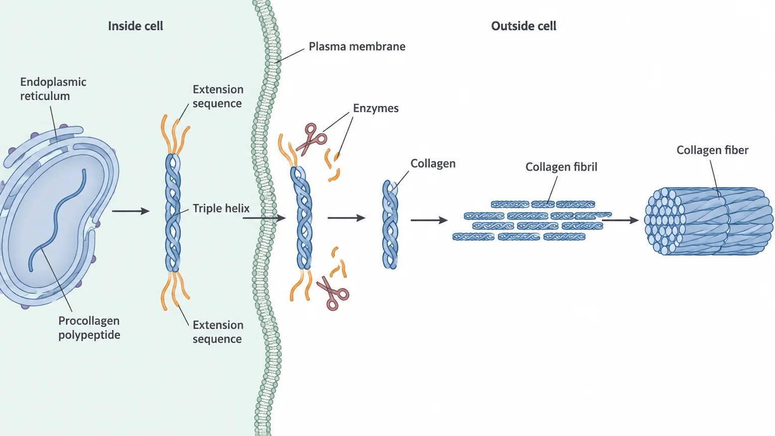
The many layers of fibrils are what give a collagen fiber its great tensile strength, the same way a rope made of many twisted strands is stronger than a single thread.
Many kinds of collagen
Animals make many types of collagen, numbered type I, type II, and so on; humans have at least 27. Each type is encoded by its own procollagen genes, and small differences in amino acid sequence change how tightly the chains and proteins pack. That produces fibers ranging from stiff to bendable, or even a meshwork instead of long fibers.
Gene regulation, the cell’s control over which genes are switched on, decides which collagen types are made where. Skin cells, for example, make types I, III, and IV but very little type II.
| Type | Made mainly in | Structure and role |
|---|---|---|
| I | Tendons, ligaments, bones, skin | Rigid, thick fibers; most abundant; most of the ECM’s tensile strength |
| II | Cartilage, discs between vertebrae | Thick but more flexible than type I; smooth joint movement |
| III | Arteries, skin, internal organs, around muscles | Thin fibers, often in a meshwork; adds elasticity |
| IV | Skin, intestine, kidneys, around capillaries | No long fibers; a meshwork that supports cell layers and filters around capillaries |
Collagen genes matter for health. Mutations in certain collagen genes cause Ehlers-Danlos syndrome, marked by very stretchy skin and overly flexible joints.
Elastin: the rubber band
Some organs, such as the lungs and blood vessels, must expand and then return to their original shape over and over. Their ECM contains elastic fibers made mainly of elastin, which is about 1000 times more flexible than collagen.
Elastin proteins are joined to each other by many covalent crosslinks, strong chemical bonds between neighboring proteins. Here is how the fiber behaves:
- Relaxed: each elastin protein curls into a compact shape.
- Stretched: the proteins pull out into straighter, more linear shapes, while the crosslinks keep them tied together.
- Released: the proteins spring back to their compact shape, and the fiber recoils.
Check yourselfHow do the structures of collagen and elastin fit their different jobs?
Collagen is many layers of fibrils bundled into thick fibers, which resist pulling and give tensile strength. Elastin is a compact protein linked by covalent crosslinks; it straightens when stretched and returns to its compact shape, which lets the fiber stretch and recoil.
Practice questions
1 of 2
A tendon must withstand strong pulling forces without tearing. Which part of its ECM mainly provides this tensile strength?
2 of 2
Skin cells make collagen types I, III, and IV but very little type II, while cartilage cells make plenty of type II. What best explains this difference?