How proteins fold into shape
A newly made polypeptide is a floppy chain. To do its job, it must fold into one precise shape, and that shape builds up in stages.
Four levels of structure
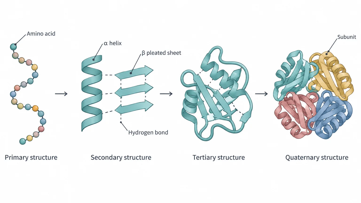
Biologists describe protein structure at four levels: primary, secondary, tertiary, and quaternary. The primary structure is the amino acid sequence. Each higher level depends on the levels below it, so changing the sequence can change the whole protein.
Secondary structure: helices and sheets
A polypeptide can bend and twist because atoms can rotate around many of the bonds in its backbone. Some stretches fold into regular, repeating patterns held in place by hydrogen bonds between backbone atoms: a hydrogen attached to a nitrogen is attracted to an oxygen that is double-bonded to a carbon. These repeating patterns are secondary structure, and there are two basic types:
- In an α helix (alpha helix), the backbone coils into a spiral, with hydrogen bonds at regular intervals holding each turn in place.
- In a β pleated sheet (beta pleated sheet), stretches of backbone lie side by side, linked by hydrogen bonds, and take on a repeating zigzag like a pleated fan.
Secondary structure affects what a protein can do. An α helix made mostly of nonpolar amino acids can anchor a protein in the oily interior of a membrane. Helices and sheets also make some proteins very strong, such as the keratin in hair and the collagen in cartilage. Stretches with neither pattern, sometimes called random coils, still have specific shapes that often matter for the protein’s function.
Tertiary and quaternary structure
As secondary structures form, side chains throughout the polypeptide interact, and the whole chain folds up on itself into a compact three-dimensional shape. That shape, helices and sheets included, is the polypeptide’s tertiary structure. For some proteins, such as ribonuclease (an enzyme that breaks down RNA), a single folded polypeptide is the finished, working protein.
Most proteins, though, are built from two or more folded polypeptides that fit together. Each polypeptide in such a protein is a protein subunit, and the subunits can be identical or different. A protein made of more than one subunit has quaternary structure. Hemoglobin, the oxygen carrier in red blood cells, has four subunits, and each binds one oxygen molecule, so one hemoglobin carries four O₂.
What holds a protein’s shape
Five kinds of chemical interactions fold a protein and keep it stable:
| Factor | What it is | Where it matters |
|---|---|---|
| Hydrogen bonds | Weak attractions between a slightly positive hydrogen and a slightly negative atom; many together add up to a strong force | Secondary structure, plus tertiary and quaternary |
| Ionic bonds and other polar interactions | Attractions between positively and negatively charged side chains, or between charged and polar side chains | Tertiary and quaternary |
| Hydrophobic effect | Nonpolar side chains cluster in the protein’s interior, away from water | Tertiary and quaternary |
| van der Waals dispersion forces | Brief, weak attractions between atoms at just the right distance; atoms too close repel, and atoms too far apart barely attract | Tertiary and quaternary |
| Disulfide bridges | Covalent S–S bonds between the sulfhydryl (–SH) groups of two cysteine side chains, in one polypeptide or between two | Stabilize tertiary and quaternary |
The first four also let different proteins bind to each other. In a protein-protein interaction, the surface of one protein fits snugly against the surface of another, held by those same forces. These precise pairings let cell processes unfold in an orderly sequence and hold larger cell structures together. Swap a few amino acids in one partner, and its surface may change enough that the two no longer fit.
Sequence determines shape
A protein’s amino acid sequence carries the information for its folding. In a key experiment, researchers treated purified ribonuclease with urea, which breaks hydrogen and ionic bonds, and β-mercaptoethanol, which breaks disulfide bridges. The enzyme unfolded, or became denatured, and stopped working.
Once the chemicals were removed, the enzyme refolded on its own and worked again, even with no other cell components present. Not every protein can do this; some need help from other proteins to fold correctly.
Domains: modular parts
Many proteins are built from modules. A domain is a region of a protein with its own distinct structure and function. During evolution, domains have been copied and reused, so one kind of domain can turn up in many different proteins, keeping the same tertiary structure and doing the same characteristic job in each.
Nuclear receptors, proteins that switch on particular genes in animal cells, show how this works. Each has separate domains for binding a hormone such as estrogen or testosterone, binding DNA, moving into the nucleus, and activating genes.
Check yourselfA mutation changes one amino acid in a polypeptide. How could that affect the protein’s secondary, tertiary, or quaternary structure?
Each level depends on the ones below it, so a single change can disrupt a local α helix, alter how the whole chain folds, or keep subunits from fitting together. In sickle cell disease, one swap in β-globin is enough to deform red blood cells.
Practice questions
1 of 4
In sickle cell disease, one amino acid in the β-globin polypeptide is replaced by a different one. Why can a single swap like this change how the protein works?
2 of 4
A protein that works in the watery fluid inside a cell folds into a compact shape. Where are most of its amino acids with nonpolar side chains likely to end up?
3 of 4
The same kind of domain, with the same three-dimensional structure and function, appears in many different proteins. What explains this?
4 of 4
Ribonuclease was unfolded with chemicals that break its hydrogen bonds, ionic bonds, and disulfide bridges. After the chemicals were removed, it refolded and worked again, with no other cell components present. What does this show?