Packing DNA into nucleosomes and loops
Stretched end to end, the DNA in one set of human chromosomes would be over 1 meter long. It has to fit inside a nucleus only about 2 to 4 micrometers (μm) across, so the DNA is hundreds of thousands of times longer than the space that holds it. Cells manage it by folding DNA in stages, with the help of proteins.
Chromosome vs. chromatin
A typical eukaryotic chromosome holds one long, linear double helix, often hundreds of millions of base pairs long. Two words describe it from different angles:
- A chromosome is one distinct unit of genetic material. A human body cell has 46 of them.
- Chromatin is the material chromosomes are made of: DNA plus the proteins packed with it (and sometimes RNA). The difference is like “a sweater” versus “wool”: you can count chromosomes, but chromatin is a substance.
Chromosomes are not frozen in one shape. They switch back and forth between tightly packed and loosely packed forms.
Nucleosomes: DNA wound on spools
The first level of packing wraps DNA around proteins called histones. Histones carry many positively charged amino acids (lysine and arginine), and DNA’s backbone is negatively charged because of its phosphates. Opposite charges attract, so DNA clings to histones.
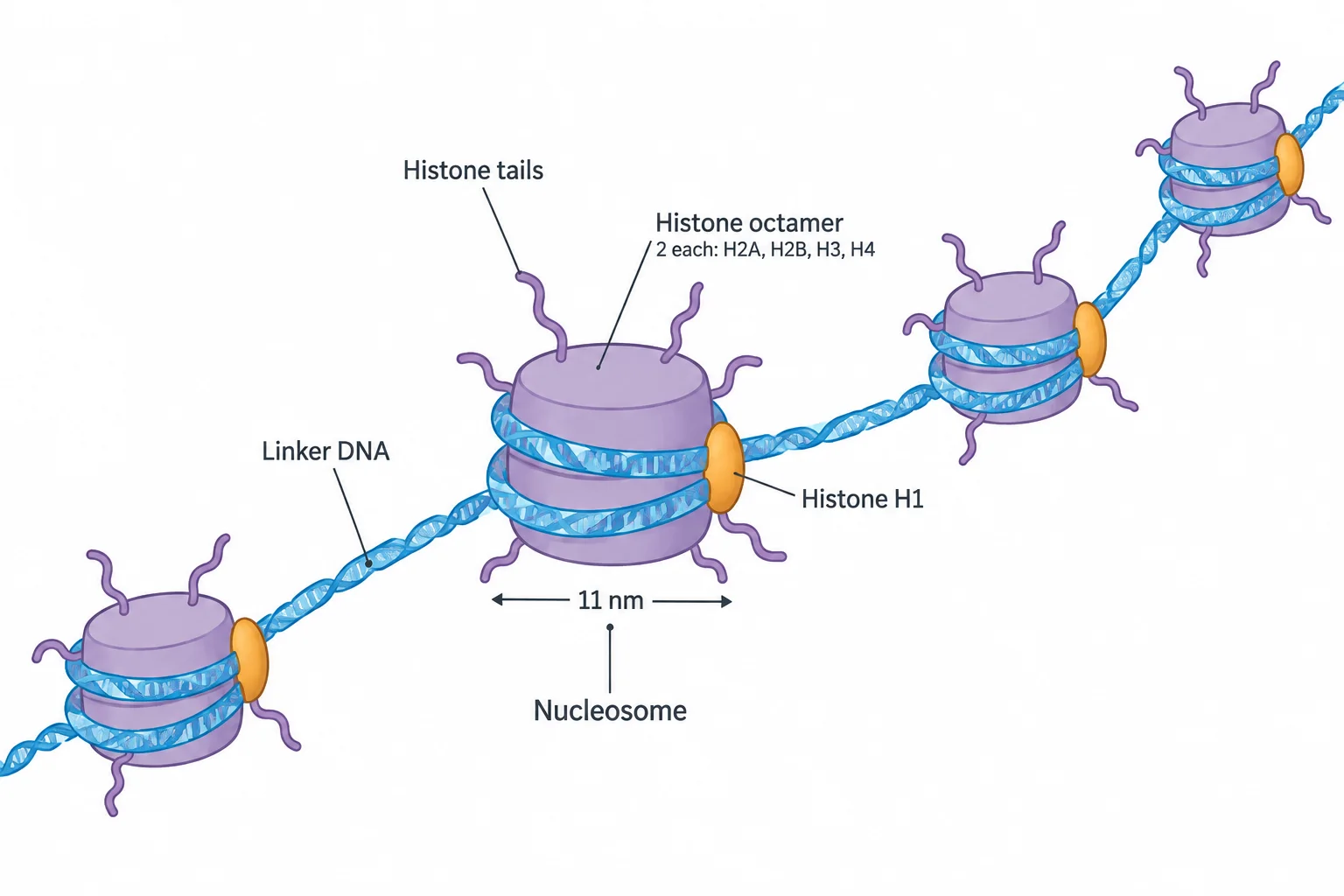
The repeating unit of chromatin is the nucleosome:
- A core of eight histones, called an octamer: two each of H2A, H2B, H3, and H4.
- About 146 to 147 base pairs of DNA wrapped around the core.
- An overall width of 11 nm at its widest.
- A tail from each histone sticking out of the core. Chemical tags added to these tails help switch genes on and off.
Neighboring nucleosomes are joined by stretches of linker DNA, 20 to 100 base pairs long depending on the species and cell type. A different histone, H1, binds to the linker region. Under the microscope the arrangement looks like beads on a string. Like thread wound on a row of spools, the DNA gets much shorter end to end without being cut: about sevenfold shorter.
Nucleosomes zigzag
Next, nucleosomes pack against one another. In the zigzag model, the linker DNA stays fairly straight and the nucleosomes alternate from side to side. As a result, each nucleosome touches not its direct neighbor but the one two positions away. In living cells, nucleosomes mostly form short zigzag clusters of three or four.
Loop domains form by loop extrusion
To pack further, stretches of chromatin fold into loops called loop domains. According to the loop extrusion model, two kinds of proteins build them:
- An SMC protein (short for structural maintenance of chromosomes) forms a pair that wraps around the DNA in a ring, making a small loop. Chromatin is then pushed, or extruded, through the ring, so the loop grows.
- CTCF (CCCTC-binding factor) binds to specific DNA sites, recognizing repeats of the sequence CCCTC. Two CTCFs sit on the DNA on either side of the growing loop and are drawn closer as it grows. When they meet, they bind each other. That stops the loop from growing and holds its base steady.
Loops can also form in other ways, for example when proteins bound to distant DNA sites grip each other. Loop extrusion, though, is a main way chromatin loops are made.
Check yourselfIn the loop extrusion model, what would happen to a loop if the two CTCFs could not bind each other?
Nothing would stop the SMC protein from passing more chromatin through its ring, so the loop would keep growing and would not be held at a set size. The SMC protein forms and enlarges the loop, while the CTCFs stop its growth and stabilize its base.
Practice questions
1 of 2
DNA wrapped around a core of eight histone proteins forms which repeating unit of chromatin?
2 of 2
What is the name for the complex of DNA and proteins that eukaryotic chromosomes are made of?