What membranes are made of
Every cell has an outer boundary that holds its contents together and screens everything trying to get in or out: the plasma membrane. Eukaryotic cells also wrap their organelles in membranes (Chapter 4). The plasma membrane and these internal membranes are all biological membranes, and they share one basic design.
Thin, flexible, and busy
A membrane is only 5–10 nm thick; thousands of them stacked together would barely match a sheet of paper. It isn’t rigid, either: membranes are flexible and somewhat fluid. Even so, they keep each cell separate from its surroundings and serve as the site of many cell activities:
- letting particular ions and molecules in and out
- dividing the cell into separate compartments
- sorting proteins to their proper destinations
- anchoring the cytoskeleton, the cell’s internal framework of protein fibers
- making energy carriers such as ATP and NADPH
- cell signaling (sending and receiving chemical messages)
- dividing the cell and its nucleus
- attaching cells to each other and to the extracellular matrix (the material surrounding animal cells)
Membrane proteins carry out most of these jobs. The lipids form the framework the proteins sit in.
The phospholipid bilayer
That framework is a double layer of fatty molecules, and water explains its shape. A water molecule is lopsided, with a slightly positive end and a slightly negative end. Molecules that also have positive and negative regions are polar, and they mix easily with water, so they’re also called hydrophilic (“water-loving”).
Molecules without such regions, such as fats and oils, are nonpolar. They don’t mix with water and cluster away from it, so they’re hydrophobic (“water-fearing”).
A phospholipid has both kinds of parts: a polar head and two long nonpolar tails. A molecule with a hydrophilic part and a hydrophobic part is amphipathic, and that split is what lets phospholipids build membranes.
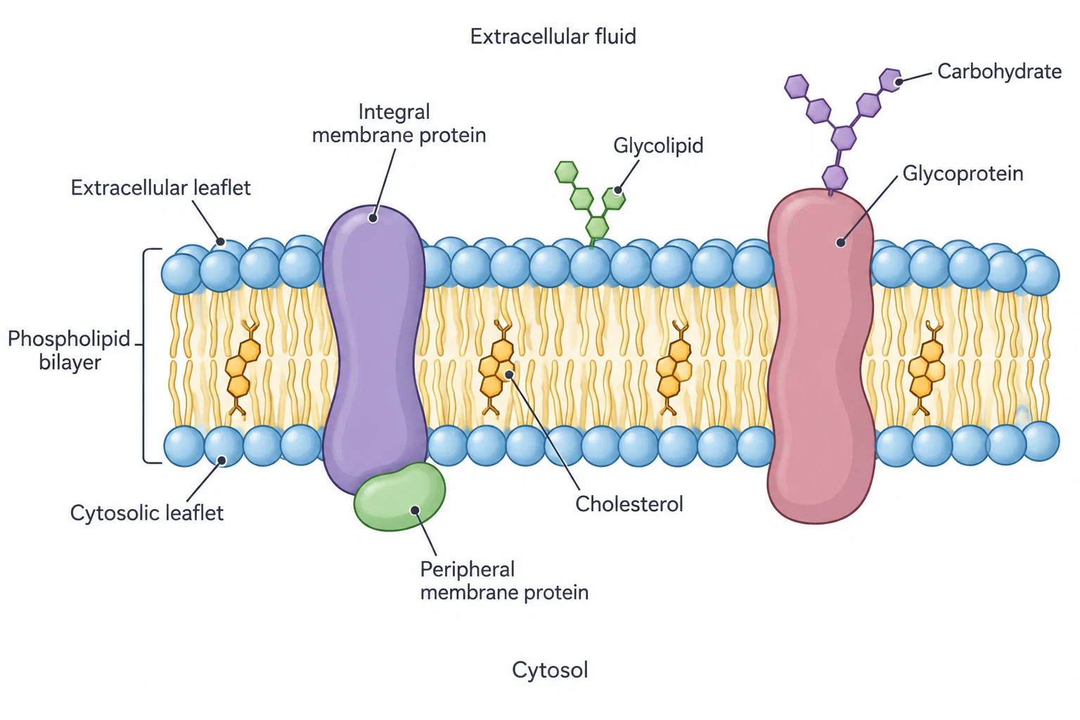
A membrane has water on both sides, so its phospholipids line up in two layers. In each layer the heads face the water, and the tails of the two layers point inward, toward each other and away from the water. This double layer is the phospholipid bilayer.
Each half of the bilayer is a leaflet. In the plasma membrane, the cytosolic leaflet faces the cytosol (the fluid inside the cell), and the extracellular leaflet faces the outside. The two leaflets aren’t identical: some kinds of lipid are more plentiful in one than in the other.
A mosaic that flows
The bilayer is only the framework. Proteins are embedded in it or attached to its surfaces, and sugar chains (carbohydrates) are attached to many of the lipids and proteins. A lipid carrying a carbohydrate is a glycolipid, and a protein carrying one is a glycoprotein (glyco- means sugar).
In the plasma membrane, these sugar chains face the outside of the cell. Glycolipids, for instance, sit mainly in the extracellular leaflet, with their carbohydrate portion sticking out into the surrounding fluid. Animal cell membranes also contain cholesterol, a lipid that tucks in among the phospholipids.
Put together, a membrane is a mosaic: a patchwork of lipid, protein, and carbohydrate molecules. It’s also fluid, because its lipids and proteins can drift past one another instead of being locked in place. This picture is the fluid-mosaic model, and it fits the membranes of all living things, from bacteria to people.
Three ways proteins attach
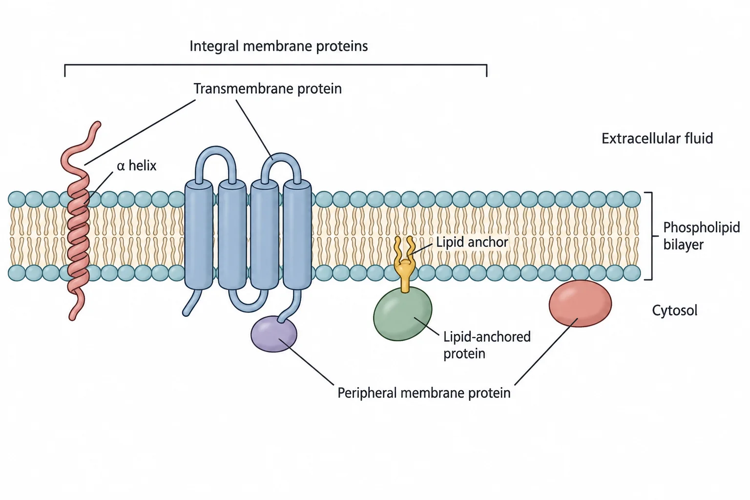
Proteins are chains of amino acids, some polar and some nonpolar. Nonpolar stretches can sit comfortably among the lipid tails, so a protein can attach in three ways, depending on how much of it enters the bilayer’s oily core:
| Type | Enters the core? | What holds it in place |
|---|---|---|
| Transmembrane protein | Yes, it spans the bilayer | One or more transmembrane segments: stretches of nonpolar amino acids, usually coiled into a spiral called an α helix, that sit among the lipid tails |
| Lipid-anchored protein | Only its lipid anchor | A lipid covalently attached (joined by a strong bond in which atoms share electrons) to one of its amino acids, with the lipid’s tails sunk into the core |
| Peripheral membrane protein | No | Weaker, noncovalent bonds, such as hydrogen and ionic bonds, to the parts of embedded proteins that stick out of the bilayer, to the polar heads of phospholipids, or to both |
Transmembrane and lipid-anchored proteins together are called integral membrane proteins, a group name rather than a fourth type: part of each one is buried in the core. They come out only if the membrane itself is dissolved with a detergent or an organic solvent, much as dish soap breaks up grease. Peripheral proteins, held only by surface attractions, can be removed while the bilayer stays intact.
Why membrane proteins matter
In bacteria, archaea, and eukaryotes (humans included), about 20–30% of genes code transmembrane proteins. Membrane proteins are also the main targets of medicines; roughly 70% of drugs act by binding one. A drug aimed at a plasma membrane protein has an advantage: it doesn’t need to get inside the cell. To test whether a drug acts on a particular membrane protein, researchers compare the protein’s function with and without the drug.
Check yourselfA protein can be washed off a membrane while the bilayer stays intact. Which of the three types is it, and what holds it in place?
It’s a peripheral membrane protein. Noncovalent bonds, such as hydrogen or ionic bonds, hold it to integral proteins or to phospholipid heads. Transmembrane and lipid-anchored proteins are integral and come out only if the membrane itself is dissolved.
Practice questions
1 of 2
Which finding, if it were true, would contradict the fluid-mosaic model of membrane structure?
2 of 2
Why do the phospholipids of a membrane form a bilayer with their tails pointing toward the middle?