X-chromosome inactivation
Imprinting sets epigenetic marks in a parent’s gametes. Other epigenetic changes start later, in the embryo, and then last into adulthood. The biggest of these silences almost an entire chromosome.
One X chromosome goes quiet
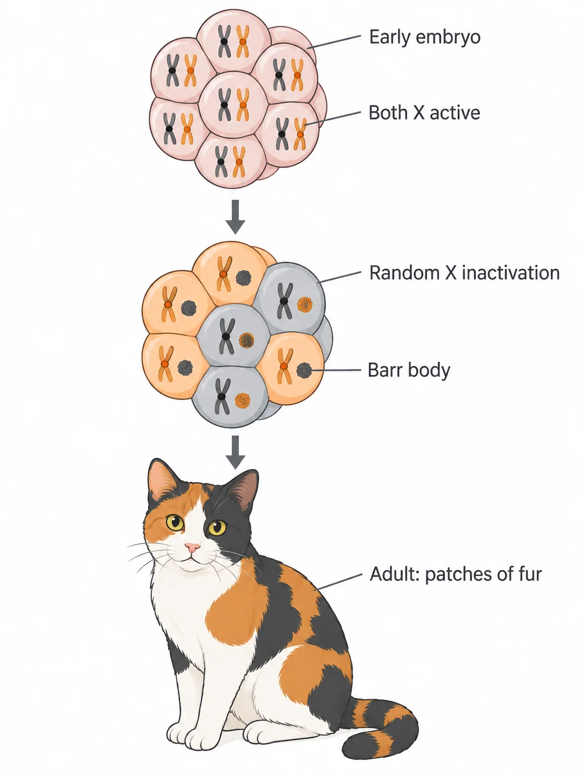
Female mammals have two X chromosomes and males have one X and one Y, so females carry two copies of every X-linked gene. Early in development, each body cell of a female embryo shuts down one of its two X chromosomes. This is X-chromosome inactivation (XCI), an epigenetic change that packs the chosen X so tightly that most of its genes are silenced.
The inactive X shows up under the microscope as a small spot at the edge of the nucleus, called a Barr body. It is far more compact than the active X in the same nucleus, so a DNA stain makes it the brightest spot there.
Why shut one down? Without XCI, females would make twice as much of every X-linked gene product as males. Silencing one X evens out the dose. Equalizing the expression of X-linked genes between the sexes is dosage compensation.
Check yourselfA DNA stain shows one X in a female cell as a small, bright spot at the edge of the nucleus. Why are so few of that X’s genes expressed?
That spot is a Barr body, packed far more tightly than the active X. The compaction keeps most of its genes from being transcribed.
A random choice that makes patchwork
Which X gets silenced is random in each cell, and the choice is permanent for that cell’s descendants.
- In the very early embryo, both X chromosomes are active in every cell.
- At an early stage, each cell randomly inactivates one X, the mother’s or the father’s.
- Every cell that descends from it inactivates the same X.
- The adult ends up with patches of tissue, each patch descended from one embryonic cell.
Calico cats show the result in their fur. An X-linked gene controls fur color, with an orange allele and a black allele, and a female heterozygous for it is calico. In a patch where the X carrying the black allele was silenced, skin cells express only orange; where the orange-carrying X was silenced, they make black. (The white patches come from a separate gene.)
The idea that random X inactivation explains this patchwork is known as the Lyon hypothesis. So a female heterozygous for any X-linked gene is a mosaic, like the mosaics of Chapter 15: her body is built from two kinds of cells, about half expressing one allele and half the other.
When mosaic tissue causes disease
Usually, half of a tissue’s cells working is enough. In hemophilia A, a recessive X-linked allele leaves the liver unable to make factor VIII, a blood-clotting protein. A heterozygous woman’s liver is a mosaic: about half her liver cells secrete factor VIII, which is plenty for normal clotting.
Rarely, a heterozygous woman has hemophilia symptoms. In humans, XCI happens when the embryo is about 10 days old, and the future liver is then only about a dozen cells. By chance, all or most of those few cells can silence the X carrying the working allele. Her adult liver then makes too little factor VIII.
Check yourselfWhy can a woman who is heterozygous for hemophilia A occasionally have symptoms of the disease?
X inactivation is random, and it happens when the future liver is only about a dozen cells. If most of those cells happen to silence the X with the working allele, too few liver cells make the clotting factor.
Counting the X chromosomes
Mammal cells count their X chromosomes and keep exactly one active; every extra X becomes a Barr body. So the number of Barr bodies is always one less than the number of X chromosomes, even in people born with an unusual number of sex chromosomes.
| Condition | Chromosomes | Barr bodies | Active X |
|---|---|---|---|
| Typical female | XX | 1 | 1 |
| Typical male | XY | 0 | 1 |
| Turner syndrome (female) | X0 | 0 | 1 |
| Triple X syndrome (female) | XXX | 2 | 1 |
| Klinefelter syndrome (male) | XXY | 1 | 1 |
People with these syndromes still show some differences, perhaps because the extra X acts before inactivation or isn’t completely silenced.
The counting depends on a short region of each X chromosome called the X inactivation center (Xic). A cell counts its Xics and inactivates every X but one. If one X in a female cell lacks its Xic, the cell counts only one, inactivates neither X, and the embryo dies.
Inside the Xic is the Xist gene. Its product is a long non-coding RNA that coats the X chromosome to be inactivated. Proteins then bind to the Xist RNA and pack the chromosome into a Barr body.
Xist keeps being expressed on the Barr body after the chromosome’s other genes fall silent, and that continued expression keeps the chromosome inactive through every cell division. When the cell divides, its Barr body is copied into two Barr bodies, one for each daughter cell.
Check yourselfHow many Barr bodies do the cells of a person with XXY chromosomes have, and how many active X chromosomes?
One Barr body and one active X. The cells count two X inactivation centers and inactivate every X but one, whether the person is male or female.
Practice questions
1 of 2
A woman is heterozygous for an X-linked gene that codes a skin enzyme. Some patches of her skin make the enzyme and other patches don’t. What explains the patches?
2 of 2
How many Barr bodies are in the body cells of a woman with triple X syndrome (XXX)?